Oosight™ system - even more accurate assessment of oocyte quality

The Oosight system makes it possible to visualise the structure of oocytes (egg cells), which is not visible under an ordinary microscope. As a result, the embryologist is able to more precisely select the best oocytes for the in vitro fertilisation procedure. This increases the chances of achieving high-quality embryos and, consequently, pregnancy and childbirth. Oosight is one of the latest technologies to increase the effectiveness of IVF.
The quality of the oocytes shows an important influence on the viability of the embryo, the subsequent development of the foetus and the course of the pregnancy. It is important both in nature and in the treatment of infertility by assisted reproduction methods. In the case of in vitro fertilisation procedures, the quality of the oocytes has a huge impact on the success of the treatment. In many cases, looking at the oocytes under the microscope with the Oosight system can have a real impact on IVF success, achieving pregnancy and having a baby.
Oocyte quality is of paramount importance for the competence of the embryo for development, implantation and subsequent fate of the foetus.
| Embryological consultation If additional questions come to mind after reading this article, remember that all Invimed patients qualified for insemination or IVF can benefit from the free embryological consultation. |
Morphological evaluation of oocytes
The ideal mature human oocyte, in terms of morphological characteristics, should be a closed, spherical structure. The oocyte must show a homogeneous and transparent cytoplasm, a single directional corpuscle of adequate size and unfragmented, an adequate thickness of the zona pellucida (ZP) and an adequate perivascular space (PVS).
Among the oocytes obtained after hormonal stimulation, there may be both normal and abnormal ones. Sometimes it happens that all the obtained oocytes have morphological defects (malformations) and are not suitable for the in vitro fertilisation procedure.
The lower quality of the oocytes is evidenced by:
-
higher incidence of structural changes,
-
presence of chromosomal abnormalities,
-
mitochondrial dysfunction,
-
displacement of oocyte components such as spindle, cortical granules or mitochondria.
Changes in oocyte morphology can result from both internal and external factors. Internal factors include a woman's age, pathological processes and genetic defects, while external factors include patients' hormonal stimulation protocols and in vitro oocyte culture conditions.
Microscopic image of abnormal oocytes
The photograph below shows morphological defects in human oocytes. 1A - centrally located cytoplasmic granules, 1B - peripherally located cytoplasmic granules; 2A, 2B - refractile bodies (arrow), 3A, 3B, presence of vacuoles; 4a,4b abnormal shape of cytoplasm; 5A, 5B - enlarged pericentriolar space; 6A, 6B - fragmentation of the first directional body (PB).
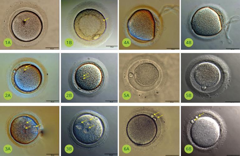
Photo 1. Microscopic images taken by R. Faundez and K. Zaręba, Invimed Warsaw, 2016.
The Oosight™ system, which is based on polarised microscopy, offers a much greater opportunity to assess oocyte quality. It allows you to see much more than is possible with traditional morphological assessment performed with a microscope without the Oosight system.
Oosight™ polarising microscopy system
The polarisation microscopy module is an additional imaging system attached to biological microscopes. The InviMed clinic in Warsaw uses the Oosight™ system from Cambridge Research & Instrumentation, Inc. It helps to determine which oocyte is best prepared for fertilisation. The system consists of the LC-PolScope™ optical-electronic module and CRi's Spindle View software, which interprets the module's camera image.
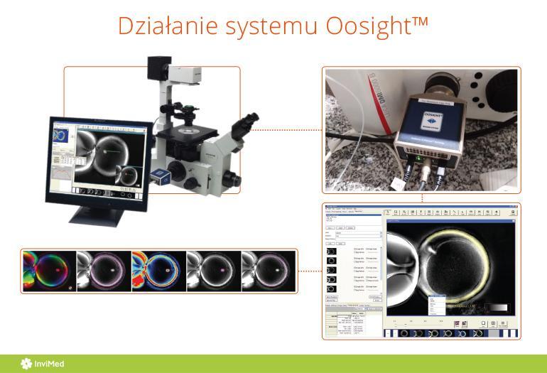
Photo.2: Operation of the Oosight system. The infographic shows a microscope with a camera and a software plug-in to display an image of an oocyte obtained using polarised light on a computer monitor.
| Advantages of Oosight Oosight provides real-time imaging. High-quality optics and unique algorithms for analysing the results guarantee accurate and reproducible data. With Oosight, the embryologist has the most objective results possible for correct oocyte classification and quality control. |
What can be seen thanks to Oosight?
The Oosight™ system allows imaging of the multilayered structure of the oocyte transparent envelope and the birefringence of the division spindle.
Transparent casing
Polarisation microscopy allows the assessment of the three-layer integrity of the oocyte's transparent envelope.
The sheath is otherwise known as the extracellular sheath, which surrounds the oocyte during its growth, maturation, ovulation, fertilisation, and in the early stages of embryonic development.
There is a correlation between the birefringence of the casing and the developmental potential of the embryo, so being able to observe the multilayered structure of the transparent casing is extremely important in assessing oocyte quality.
Dividing spindle
The division spindle is a very important cellular structure that enables the precise division of genetic material (chromosomes) - the correct maturation of the nuclear egg cell.
Polarisation microscopy allows visualisation of the spindle and a more accurate analysis of its shape, size and birefringence, making it easier for embryologists to predict possible anomalies in chromosome segregation. Lack of spindle visualisation at a specific time of oocyte maturation is significantly correlated with poorer viability, poorer development and lower embryo quality.
Oocyte image from under the microscope with the Oosight system
The operation of the Oosight system is non-invasive. In polarisation microscopy, neither exogenous dyes are used for imaging, nor are cells exposed to light that is harmful to their structures. Instead, the optical property of cell structures, i.e. birefringence (the ability to double refract light), is exploited.
The images below show an image of an oocyte obtained with a microscope equipped with the Oosight system. 1) Mature oocyte with normal morphology, showing the zona pellucida (ZP), primordium (PB) and division spindle (WP) in the correct location; 2) Mature oocyte, spindle in the wrong location; 3) Mature oocyte, absence of primordium.
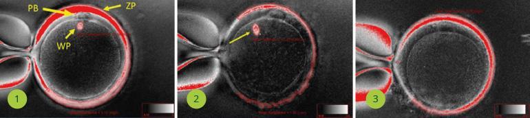
Photo 3. Photographs from the microscope with the Oosight system taken by S. Dabrowski, B. Szkoda, Invimed Warsaw.
| Oosight in IVF clinics Oosight is an effective new tool used in top IVF clinics around the world for ICSI/IMSI in vitro fertilisation procedures, as well as for oocyte selection for the vitrification procedure. |
Oosight a ICSI/IMSI
Oosight also provides increased accuracy in the execution of procedures ICSI and IMSIThis involves injecting a pre-selected sperm directly into the egg. The Oosight-enabled determination of the presence and exact positioning of the spindle during ICSI/IMSI results in an even higher success rate for these procedures. Studies suggest that positioning the spindle at the sixth or twelfth hour during sperm injection into the egg leads to less embryo fragmentation, i.e. better embryo quality and higher implantation rates.
Oosight versus oocyte vitrification and thawing
The Oosight system also allows monitoring of the effects of oocyte cryopreservation (vitrification). The division spindle is temperature-sensitive and highly susceptible to damage during cryopreservation. Using the Oosight system, it is possible to assess the integrity of the spindle after thawing.
Find out more about vitrification, the slow freezing of embryos and oocytes >>.
***
Article elaborated: Dr Ricardo Faundez, chief director of embryology at Invimed infertility clinics. Member of prestigious scientific societies, including ESHRE. One of the first embryologists in Poland to deal with assisted human reproduction by in vitro method.
The medical information presented should be considered as general guidelines and does not replace the individual judgement of the doctor regarding the medical management of each patient. The doctor, after a thorough examination of the patient's condition, determines the extent and frequency of diagnostic tests and/or therapeutic procedures, taking into account specific medical indications. All medical decisions are made in full consultation with the patient.
Author of the article
Invimed editorial team - we serve patients by solving their fertility problems. We use world medical knowledge, state-of-the-art technology and treatment methods. We are here to make dreams of parenthood come true. The smiles on the faces of happy parents give meaning to our work.
See all articles →