Sonovaginography at the Invimed clinic in Wrocław

Sonovaginography is an advanced ultrasound examination using gel contrast for patients with suspected endometriosis. This examination allows the evaluation of anatomical structures of the pelvis minor that cannot be detected by routine ultrasound. Sonovaginography is a key element in the diagnosis of endometriosis, a disease that is difficult to detect without the use of specialised imaging methods. Endometriosis ultrasound with gel contrast is performed in the clinic Invimed in Wroclawwhere experienced specialists provide the highest standard of diagnostics.
Diagnosis of endometriosis - what is sonovaginography
Endometriosis is a silent, extremely insidious disease that can affect up to one million Polish women, affecting their health and quality of life. The condition is characterised by the presence of endometrial cells outside the uterine cavity, which causes a number of discomforts. Patients with endometriosis complain of very severe chronic pain, heavy periods, problems with urination or bowel movements, constipation, diarrhoea, flatulence, among others. Untreated endometriosis can cause infertility. It is estimated that up to 50% women unable to get pregnant suffer from the disease.
Endometriosis is described as a 'mystery' disease because it is difficult to diagnose. Some women wait several years or more for a proper diagnosis. Meanwhile, as the years pass, the disease progresses, leading to many complications. Therefore, proper diagnosis of endometriosis is extremely important!
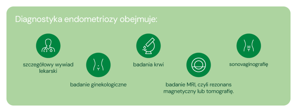
The basis for diagnosing the disease is a visit to a specialist who has the necessary knowledge and experience.
Sonovaginography is specialised ultrasound examination of endometriosis performed with the use of gel contrast. The examination is performed when superficial and deeply infiltrating endometriosis foci are suspected. Sonovaginography is performed using a vaginal probe. Thanks to the use of a special gel that acts as a contrast agent, a more accurate and precise assessment of organs including the vagina, ligament, kidneys, bladder or ureters is possible than during a traditional ultrasound examination.
What is sonovaginography? Key stages of the examination
Ultrasound examination of endometriosis with gel contrast can detect foci of superficial and deeply infiltrating endometriosis.
Sonovaginography - what it looks like course of the test?
-
Sonovaginography is performed using a vaginal probe. Prior to the examination, the doctor uses a special catheter to introduce a gel into the vagina, which acts as a contrast agent. This allows him or her to identify endometriosis on ultrasound at a much more sensitive level than during a standard ultrasound.
-
In the first stage, the kidneys are examined to rule out possible urinary stasis, which may be caused by ureteral stricture in the course of endometriosis. The doctor assesses the reproductive organ, i.e. the cervix, the uterine body and the ovaries. In addition, the mobility of the ovaries and uterus is assessed.
-
This can be followed by a thorough assessment of the area around the anterior vaginal wall, bladder, posterior vaginal wall, rectovaginal septum and rectum for foci of deeply infiltrating endometriosis.
Endometriosis ultrasound with contrast gel examination takes longer than a standard gynaecological examination. Because deeper structures are assessed, discomfort may be experienced.
Who should perform sonovaginography and when?
Sonovaginography is recommended for patients with suspected deep infiltrating endometriosis. This is the most severe and advanced form of this disease.
What symptoms could indicate a disease?


Ultrasound for endometriosis is performed on the indication of the doctor. Each case requires a consultation and determination of the diagnostic process with a specialist.
Preparing for endometriosis ultrasound - what do you need to know?
Many patients wonder how to prepare for an endometriosis ultrasound? Meanwhile, sonovaginography is an examination that does not require complicated preparations. The key element, is to ensure that the bladder is adequately filled, which allows for optimal image quality.
Endometriosis ultrasound with the use of contrast can be carried out in any phase of the cycle, including during menstruation. However, for the comfort of the patient and for a more precise evaluation of the endometrium, it is recommended to perform the examination in the first phase of the cycle. At this time, the diagnostic conditions are particularly favourable, which may contribute to a more accurate assessment of the changes.
How much does an endometriosis ultrasound cost?
Sonovaginography is an advanced ultrasound examination that requires not only highly specialised equipment, but above all the knowledge and experience of the person performing the examination. Skilful use of advanced ultrasound technology is key, allowing a precise assessment of lesions and a reliable diagnosis to be made.
Only such an approach guarantees a reliable assessment of the patient's condition and enables the implementation of appropriate treatment.
At the Invimed clinic in Wrocław, the price of sonovaginography examination is PLN 600.
Where to perform endometriosis ultrasound in Wrocław?
Specialist ultrasound for endometriosis is performed at the Invimed clinic in Wrocław. Sonovaginography is performed by highly qualified specialists with extensive experience in the diagnosis of endometriosis. Thanks to their knowledge and precision, patients can be sure that the results will be reliable and credible.
To make an appointment for endometriosis diagnosis in Wrocław, please:
-
call the helpline on 500 900 888
-
complete and submit the form on this page
-
send e-mail to: invimed@invimed.pl
-
sign up at the reception desk at the Invimed clinic in Wroclaw
Bet on precision and experience - make an appointment for sonovaginography at Invimed in Wrocław now!
The medical information presented should be considered as general guidelines and does not replace the individual judgement of the doctor regarding the medical management of each patient. The doctor, after a thorough examination of the patient's condition, determines the extent and frequency of diagnostic tests and/or therapeutic procedures, taking into account specific medical indications. All medical decisions are made in full consultation with the patient.
Author of the article
Invimed editorial team - we serve patients by solving their fertility problems. We use world medical knowledge, state-of-the-art technology and treatment methods. We are here to make dreams of parenthood come true. The smiles on the faces of happy parents give meaning to our work.
See all articles →